Lumps and bumps of the upper or lower eyelids are fairly common occurrences.
Besides those attributable to infection (stye) and blockage of the oil glands (chalazion), which I have written about (see section on Lumps and Bumps of the Eyelids- Infection of the Eyelids), lumps and bumps that are essentially growths on the eyelids are just as important and merits more than just a cursory mention in any decent website for the benefit of patients.
The reason for this is because such lesions of the eyelids may very well turn out to be malignant, i.e., Cancers of the eyelids, and warrants utmost attention to prevent further morbidity and further devastating sequelae.
What are the warning signs that a tumour may be malignant?
These questions are frequently asked, and I shall attempt to shed light on this important issue.
1. The tumour grows rapidly in size
2. The tumour bleeds
3. The tumour thickens
4. The tumour edges are irregular
Why should I see an Eyelid specialist for this condition?
Besides the possibility that the lesion may turn out to be malignancies if they manifested the four features as mentioned above, sometimes self-monitoring might not suffice.
Even if looked at in the mirror or with a magnifying glass by relatives, this might not be sufficient to show up patterns that may suggest malignant transformation.
At our practice, Dr Chuah will examine your lesion with a slit lamp, where the image is magnified many times more than even with a magnifier.
In addition, Dr Chuah will record and project what he sees on a monitor, so that patients will be able to see as much as is helpful to aid all concerned ( including their relatives) come to a decision on what should be done.
What happens after the consult?
A consultation does not necessarily imply that surgery needs to be undertaken.
Patients have the options of observing the lesion, other non-surgical manoeuvres (such as warm compresses for the infected styes-see section on infection of the eyelids), and lastly, excision.

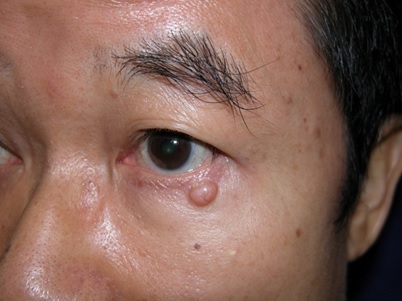
The tumour in the example shown here increased in thickness and grew rapidly over two months.
The patient underwent excision biopsy and reconstruction with the biopsy result of a basal cell carcinoma- the most common form of eyelid cancer. (We apologise for not showing the “after” picture, in keeping with MOH rules. Our main aim is to educate and increase patients’ awareness).

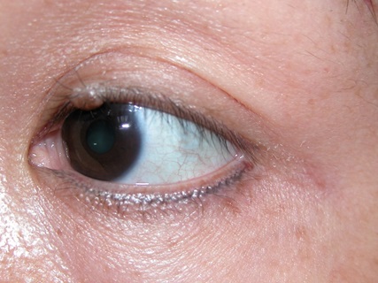
This patient has a left upper eyelid lesion that is benign.
It is a naevus that the patient says she has had since childhood.
A naevus can be congenital or acquired, with the acquired variety appearing between 4-15 years of edge. Rarely (less than 1%), these lesions undergo malignant transformation to the malignant version known as malignant melanoma, and for this reason, they should be observed by an eye doctor.

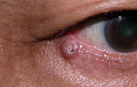
The above lesion is a basal cell carcinoma- it illustrates the point that when a lesion has irregular edges, our index of suspicion should be raised.
